
Many patients have questions about the use of rib cartilage in rhinoplasty, particularly when deciding between their own rib cartilage (autologous costal cartilage) and donor rib cartilage (cadaveric or allogeneic costal cartilage). Understanding the differences between these materials is important when considering primary rhinoplasty or revision rhinoplasty. In this article, Dr. Gorn explains their preparation, surgical applications, advantages, limitations, and potential complications to help patients make more informed decisions.
Primary and Revision Rhinoplasty Using Your Own Rib Cartilage (Autologous Costal Cartilage | Rib Cartilage Rhinoplasty)
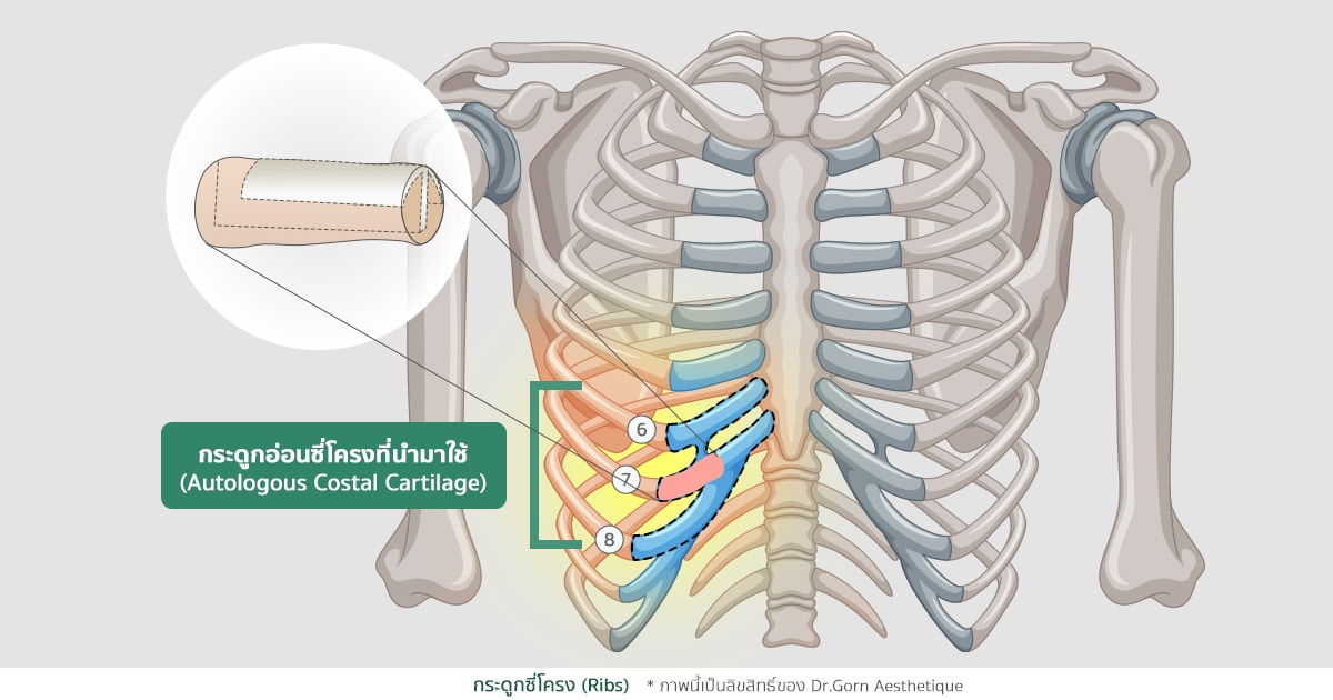
Autologous costal cartilage rhinoplasty involves harvesting cartilage from the patient's own rib cage and using it to reconstruct or support the nasal framework. The original surgical approach described here commonly considers the sixth, seventh, or eighth rib cartilage on the right side of the chest. The amount harvested depends on the structural requirements and the surgeon's judgment.
Before surgery, the surgeon may recommend a chest CT scan to assess the available rib cartilage. The original article describes four main objectives of this examination:
- To measure the diameter, length, and available cartilage portion of individual ribs.
- To evaluate the quality of the rib cartilage, including the degree of calcification that might affect graft harvesting and shaping.
- To determine whether sufficient suitable cartilage is available for the planned nasal augmentation or structural reconstruction.
- To identify relevant abnormalities of the chest or lungs that could influence surgical planning and anesthesia assessment.
After reviewing the imaging results, the surgeon determines whether the patient's rib cartilage is suitable for the planned nasal reconstruction. Important considerations include cartilage quality, the available quantity, and whether the graft can provide adequate support for the specific nasal concerns. Although chest CT can be helpful in selected cases, it is not universally required before every rib cartilage rhinoplasty procedure.
Other preoperative examinations may include a physical examination, blood tests, chest X-ray, coagulation studies (coagulogram), electrocardiography (ECG), and additional investigations depending on the patient's age, medical history, and health status. The anesthesiologist evaluates the patient's suitability for anesthesia and helps identify potential perioperative risks. Not every patient requires every investigation listed here.
How Is Rib Cartilage Harvested for Rhinoplasty?
The surgeon makes an incision over the selected rib cartilage, commonly measuring approximately 3–4 cm in the approach described in the original article. A longer incision may be necessary for patients with thicker skin or greater subcutaneous tissue to provide adequate surgical exposure.
Rib cartilage harvesting is commonly performed under general anesthesia, particularly when combined with complex structural rhinoplasty. In female patients, the incision may be placed beneath the breast to help conceal the scar. Patients who have previously undergone breast augmentation with silicone implants should inform their surgeon before surgery so the incision and harvesting approach can be planned with consideration for the breast implants. The final incision position and anesthesia technique depend on individual circumstances.

Postoperative Wound Care After Autologous Rib Cartilage Harvesting
Postoperative wound care is a common concern for patients considering rhinoplasty using their own rib cartilage. Many patients want to know whether the chest incision is difficult to care for and how soon they can resume normal activities.
In uncomplicated cases, patients are generally encouraged to begin gentle movement and walking according to their surgeon's instructions. The original article recommends avoiding heavy lifting, direct impact to the chest, and strenuous physical activities for approximately 30 days after surgery. Actual restrictions depend on the procedure, healing progress, and the treating surgeon's recommendations.
The postoperative wound-care schedule described in the original article includes:
- Days 1–2: Clean and care for the nasal surgical wound as instructed. Leave the rib cartilage donor-site dressing in place unless the medical team advises otherwise.
- Day 3: The chest incision dressing may be opened or changed by the medical team according to the wound-care plan.
- After the dressing is opened: The original protocol describes cleaning the wound with normal saline, applying a scar-care preparation, allowing the area to dry, and using a prescribed antibiotic ointment in the morning and evening until the wound heals sufficiently. These products should only be used as directed by the surgeon, because treatment for an open incision differs from scar care after wound closure.
- Suture removal: Sutures may be removed approximately 7–14 days after surgery, depending on the type of sutures and individual wound healing.
- Physical activity: Avoid strenuous exercise for approximately 30 days, or for the period recommended by the surgeon.
- Scar management: Once the incision has fully closed and the surgeon confirms that it is appropriate, a suitable scar-care gel may be applied in a thin layer as instructed.
- Postoperative discomfort: Pain or tenderness at the rib harvesting site may occur during the first 1–2 weeks and usually improves progressively. Severe or worsening pain, breathing difficulty, fever, or abnormal wound drainage requires prompt medical assessment.
Note: In selected structural rhinoplasty procedures, the surgeon may preserve unused portions of harvested rib cartilage for potential future reconstruction. The original article describes storing some remaining cartilage in deeper tissue near the harvesting incision or beneath the scalp in the lateral occipital region. This is a surgical technique used in selected circumstances and should not be assumed to be routine or appropriate for every patient.
Advantages and Disadvantages of Autologous Rib Cartilage in Primary and Revision Rhinoplasty
Advantages
- Adequate Cartilage Supply and Strong Structural Support: Autologous rib cartilage can provide a relatively large quantity of cartilage with sufficient strength for complex structural rhinoplasty and revision procedures, particularly in patients with the following concerns:
-
- Short and Upturned Nose: Patients requiring structural support to lengthen a short or upturned nose may benefit from rib cartilage grafting.
- Deviated Nasal Septum or Mid-Vault Collapse: Reconstruction may require strong cartilage grafts in sufficient quantities to correct structural deformities.
- Thick or Oily Nasal Skin: Patients with a thick, less flexible skin-soft tissue envelope or significant scarring from previous operations may require additional structural support.
- Complex Nasal Deformities: Rib cartilage may be useful when extensive structural correction is necessary.
- Revision Rhinoplasty: Particularly in patients who have undergone previous silicone implant rhinoplasty or whose septal or ear cartilage has already been harvested or used.
- Use of the Patient's Own Tissue: Autologous cartilage is biologically compatible with the patient and avoids the need for donated graft material. Nevertheless, infection and other graft-related complications remain possible.
- Availability of Rib Perichondrium: The patient's own rib perichondrium may be used as a tissue covering or for selected reconstructive purposes, including cases with thin nasal tip skin.
Disadvantages
- Longer Operating Time: Harvesting rib cartilage adds an additional surgical step and can increase the duration of rhinoplasty.
- Chest Wall Scar: An incision is required at the donor site, often on the right side of the chest. Scar appearance generally changes over time and depends on individual healing characteristics.
- Cartilage Rigidity and Calcification: Rib cartilage is primarily hyaline cartilage and may become increasingly calcified with age. Depending on the degree of calcification and graft design, it may feel firmer than elastic cartilage obtained from the ear.
- Donor-Site Surgical Risks: Rib cartilage harvesting is a specialized operation and may cause complications, including:
- Postoperative pain at the harvesting site.
- Postoperative bleeding or hematoma.
- Graft fracture or difficulty shaping cartilage with substantial calcification.
- Injury to the pleura that may result in pneumothorax (air entering the space around the lung).
Primary and Revision Rhinoplasty Using Donor Rib Cartilage (Cadaveric Costal Cartilage / Allograft Cartilage)
Donor rib cartilage, also called cadaveric costal cartilage or homologous/allogeneic costal cartilage, is another graft option for nasal augmentation and reconstruction. It may be considered when the patient cannot use their own rib cartilage or wishes to avoid additional chest wall surgery, but still requires a relatively large amount of cartilage for structural rhinoplasty or revision nasal reconstruction.
Donor cartilage undergoes tissue processing and preparation before clinical use. Processing methods vary by tissue bank, manufacturer, and product. The original article lists the following methods:
- Hydroxide oxidation.
- Demineralization.
- Lyophilization.
- Gamma irradiation, including irradiated homologous costal cartilage (IHCC).
- Freeze-drying processes.
Depending on the preparation method, donor cartilage may contain nonviable chondrocytes within a structural matrix composed mainly of collagen and proteoglycans. Not every donor cartilage product undergoes all the processes listed above. Appropriate donor screening, processing, storage, and traceability are important considerations when selecting graft material.
When May Donor Rib Cartilage Be Considered for Rhinoplasty?
Donor rib cartilage may be considered for patients who have limitations affecting the use of their own rib cartilage, including:
- Patients who wish to avoid an additional rib harvesting procedure or for whom harvesting presents additional medical or anesthetic concerns. Using donor cartilage does not automatically eliminate the need for general anesthesia during rhinoplasty.
- Older patients in whom the suitability of harvesting autologous rib cartilage requires careful assessment.
- Patients with multiple underlying medical conditions that could increase the risks of donor-site surgery.
- Patients whose own rib cartilage is unsuitable because of poor quality, extensive calcification, or complete ossification.
Advantages and Disadvantages of Donor Rib Cartilage in Rhinoplasty
Advantages
- Potentially Shorter Operating Time: Because donor cartilage does not require harvesting from the patient's chest, the overall operation may be shorter than rhinoplasty involving autologous rib cartilage harvesting.
- Anesthesia Flexibility in Selected Cases: Some procedures may be performed using local anesthesia with or without sedation, depending on surgical complexity and patient suitability. Complex revision cases may still require general anesthesia.
- No Chest Wall Donor-Site Incision: Avoiding rib cartilage harvesting eliminates the additional chest scar and the complications specifically associated with the harvesting procedure.
Disadvantages
- Potential Graft Infection or Other Complications: The original article describes a concern that donor cartilage may have lower resistance to infection than autologous cartilage. However, comparative studies have not consistently demonstrated a statistically significant difference in infection rates. Risks depend on factors such as tissue processing, patient characteristics, and surgical technique.
- Changes in Material Characteristics: Sterilization and tissue processing may affect the mechanical properties of certain donor cartilage preparations, potentially increasing brittleness or influencing handling characteristics.
- Limited Use of Donor Perichondrium: Donor cartilage products may not provide suitable perichondrial tissue for use as a covering in patients with thin nasal tip skin. The suitability of additional tissue grafts should be assessed separately.
Research Findings on Complications of Rib Cartilage Grafts in Rhinoplasty
Several clinical studies and systematic reviews have investigated the outcomes and complications associated with rib cartilage grafting in rhinoplasty. The following findings summarize the two published meta-analyses referenced in the original article. The reported percentages represent findings from the studies included in those reviews and should not be interpreted as guaranteed outcomes for individual patients.
- Meta-analysis [1]: Autologous Rib Cartilage Rhinoplasty Complications. The original article describes an analysis involving 10 studies published or identified within the period 1946–2013, including 491 patients who underwent rhinoplasty using autologous rib cartilage. Follow-up periods ranged from approximately 1.5 to 8 years. The original article reports the following complication ranges:
-
- Cartilage warping: 1–5%.
- Graft resorption and contour irregularity: 1–2%.
- Infection: 2–5%.
- Graft displacement: approximately 1%.
- Revision surgery: 2–12%.
- Meta-analysis [2]: Comparison of Autologous vs Homologous Costal Cartilage Grafts in Dorsal Augmentation Rhinoplasty. The original article describes a systematic review involving research published between 1990 and 2017, with a reported total of 1,041 rhinoplasty cases across 11 countries. The groups described include 741 autologous rib cartilage cases and 293 donor rib cartilage cases. The donor cartilage groups included irradiated homologous costal cartilage (IHCC) and Tutoplast-processed cartilage, particularly for en bloc dorsal onlay grafts. The group figures are reproduced as stated in the original article and do not sum to its stated total.
-
- IHCC donor cartilage: 153 cases, with a mean follow-up of 31.2 months (reported range: 5.4–57 months).
- Tutoplast donor cartilage: 140 cases, with a mean follow-up of 18.7 months (reported range: 0.0–79.1 months).
- Autologous costal cartilage: 741 cases, with a mean follow-up of 23.2 months (reported range: 13.8–32.7 months).
The original article reports the following overall complication estimates:
-
- Graft resorption: 2% (0–2%).
- Infection: 2% (0–4%).
- Cartilage warping: 5% (3–9%).
- Contour irregularity: 1% (0–3%).
- Revision surgery: 5% (2–9%).
Note: The original article states that no statistically significant differences in the reported complication rates were found between the two donor cartilage preparation groups and the autologous cartilage group. This does not establish that the materials are identical in all respects or that their risks are equivalent for every patient. Study design, sample size, surgical techniques, follow-up periods, and graft processing methods should be considered when interpreting these findings.
[1] Reference: Autologous Rib Cartilage Rhinoplasty Complications. JAMA Facial Plastic Surgery. January/February 2015;17(1).
[2] Reference: Comparison of Autologous vs Homologous Costal Cartilage Grafts in Dorsal Augmentation Rhinoplasty: A Systematic Review and Meta-analysis. JAMA Otolaryngology–Head & Neck Surgery. 2020;146(4):347–354.