
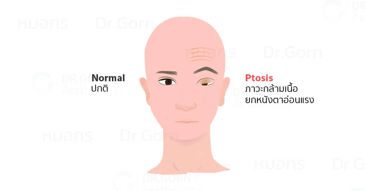
Upper Eyelid Ptosis (Blepharoptosis)
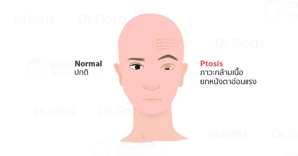
Ptosis can affect people of any age or sex. It may involve one or both upper eyelids, be present from birth, or develop later in life. The degree of drooping may also differ between the two eyes. Some people notice more pronounced eyelid drooping after botulinum toxin injections to the forehead. Others may experience worsening drooping or greater eyelid crease asymmetry after conventional double eyelid surgery. Previously unrecognised ptosis may be one contributing factor.
Identifying ptosis before surgery is important. It is often confused with dermatochalasis, which refers to excess or loose eyelid skin. These conditions require different assessments and may need different treatment approaches.
What Is Ptosis?
Ptosis describes an abnormally low position of the upper eyelid. It can result from a problem in any part of the mechanism responsible for lifting the eyelid, including the midbrain, the third cranial nerve, the neuromuscular junction, the levator palpebrae superioris muscle, the levator aponeurosis, or Müller’s muscle.
The levator aponeurosis connects the levator muscle to the eyelid’s tarsal plate. Identifying which part of the eyelid lifting mechanism is affected is essential when selecting an appropriate treatment.
How Upper Eyelid Elevation Is Controlled
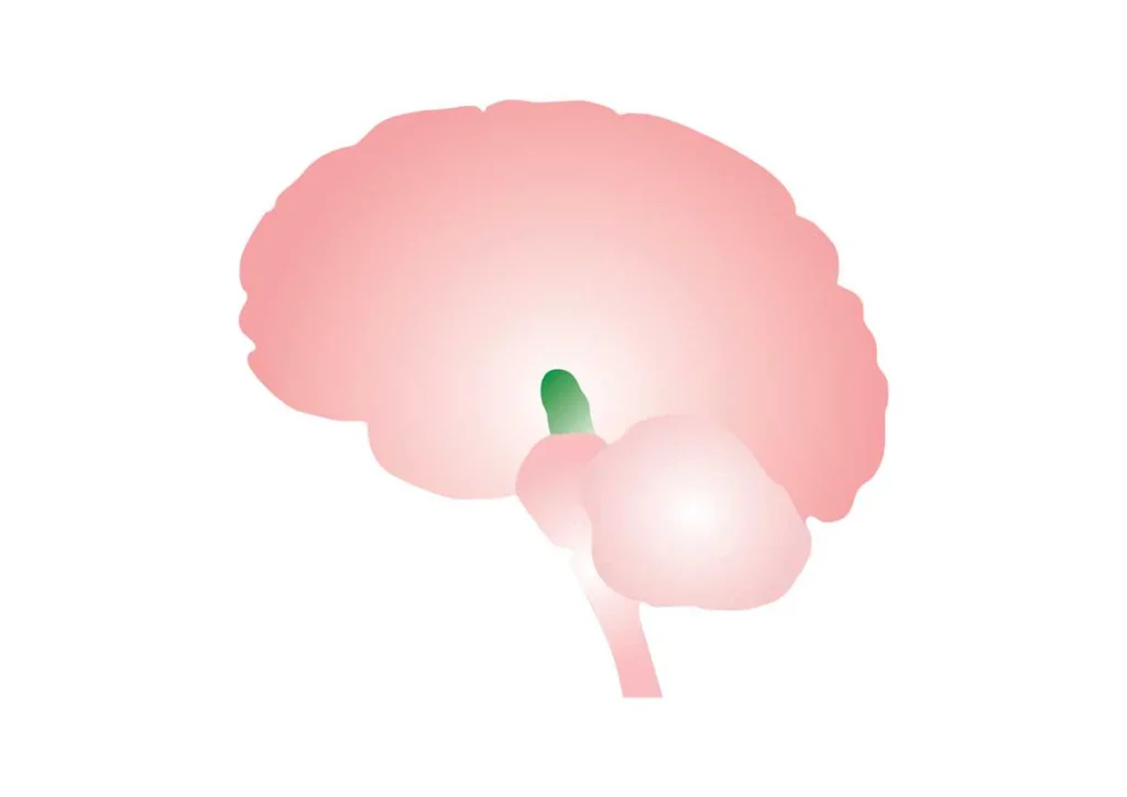
Midbrain
The middle part of the brain involved in controlling upper eyelid elevation.
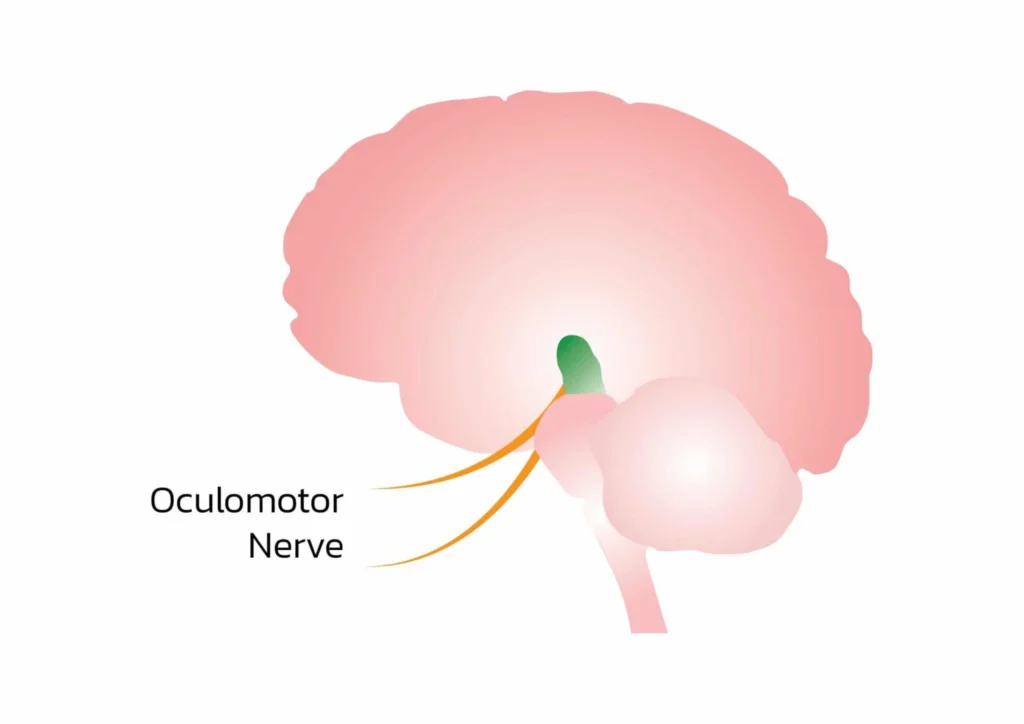
Third Cranial Nerve
The oculomotor nerve, also known as cranial nerve III.
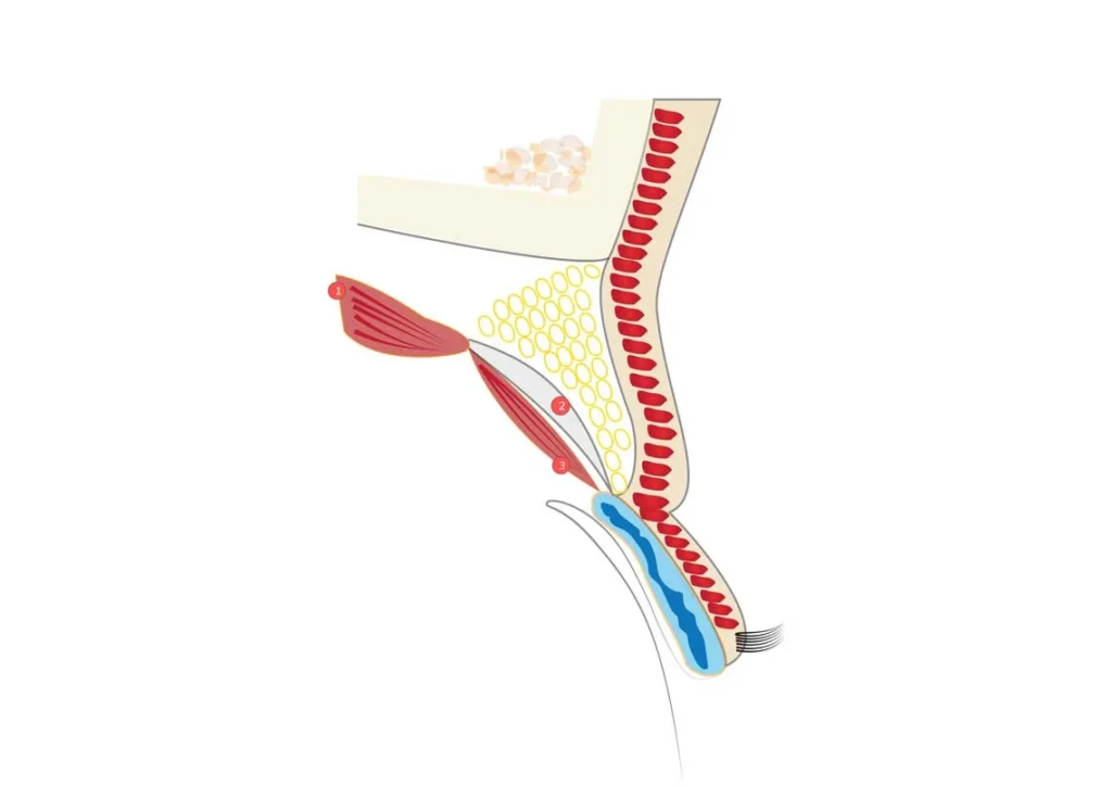
Levator Complex
The structures involved in lifting the upper eyelid.
- When we open our eyes, signals from the brain travel through the third cranial nerve to the levator palpebrae superioris muscle.
- The levator muscle contracts, lifting the upper eyelid.
- When we close our eyes, levator activity decreases, allowing the upper eyelid to descend as the eyelid closing muscles act.
Structures Involved in Upper Eyelid Elevation
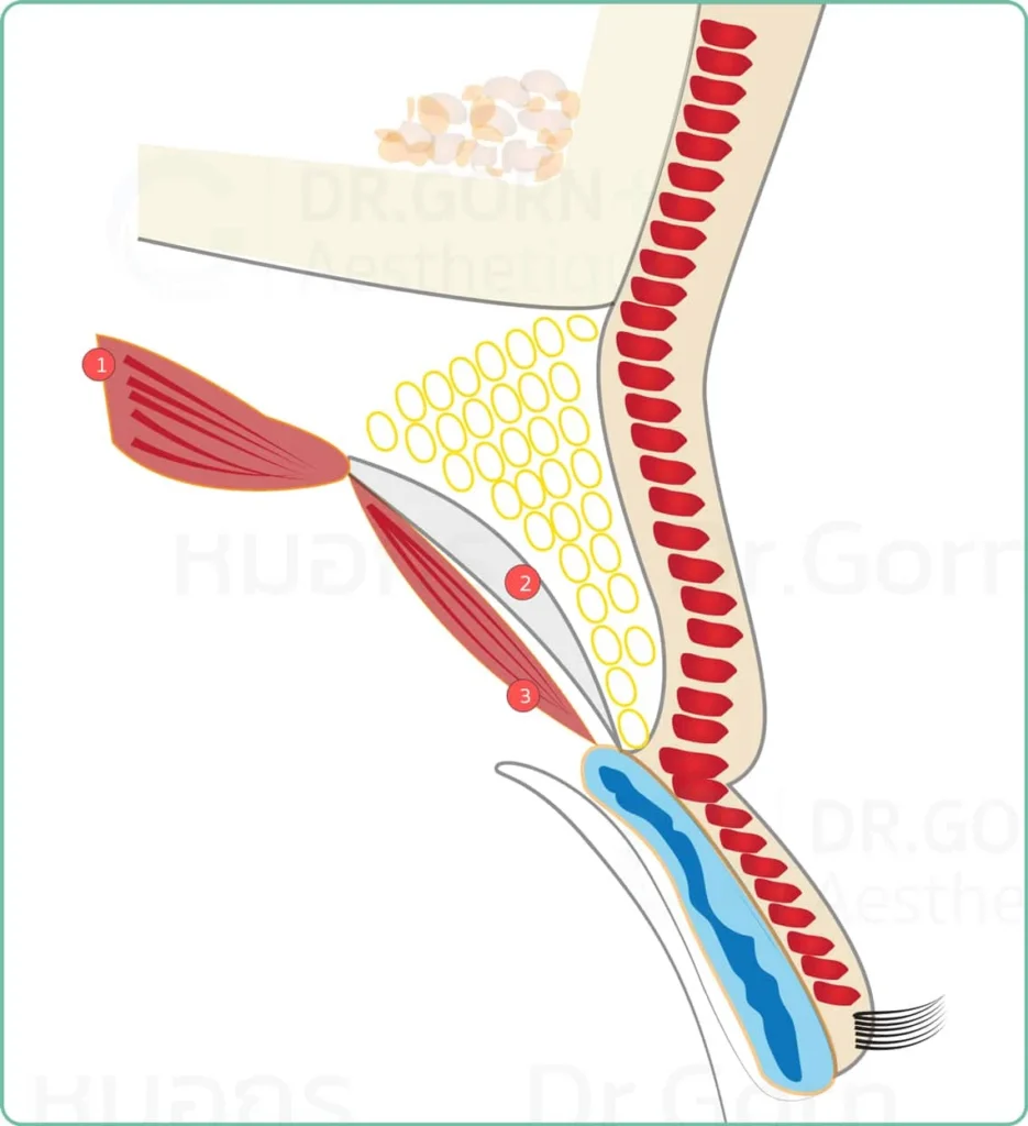
Three Main Components
- Levator palpebrae superioris muscle.
- Levator aponeurosis.
- Müller’s muscle.
How Can You Recognise Possible Ptosis?
One initial observation is the position of the upper eyelid while looking straight ahead with the face relaxed. Recent botulinum toxin injections around the eyes or forehead can affect this assessment and should be reported to the doctor.
The original article describes the upper eyelid as normally covering approximately 1 mm of the upper cornea in younger people and approximately 2 mm in older people. Greater coverage may suggest ptosis, but these measurements alone do not establish a diagnosis. A doctor must assess eyelid position, lifting function, and the overall eye condition.
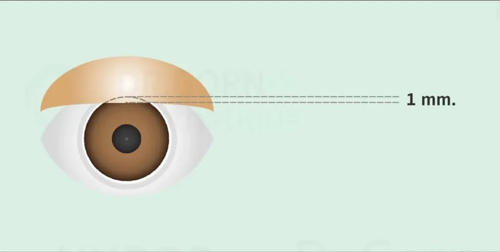
Reference Illustration for Younger People
The upper eyelid covers approximately 1 mm of the upper cornea, as described in the original article.
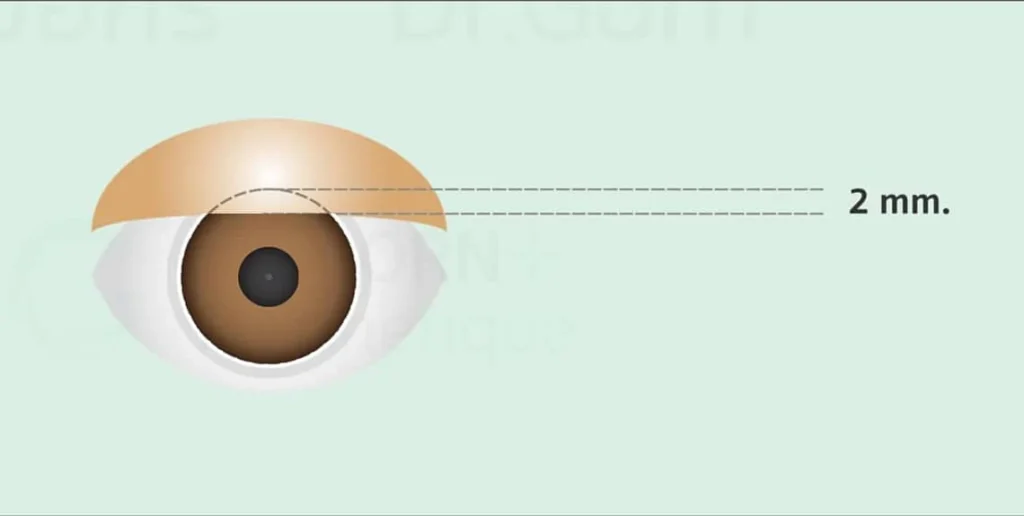
Reference Illustration for Older People
The upper eyelid covers approximately 2 mm of the upper cornea, as described in the original article.
- Ptosis may be suspected when the upper eyelid sits lower than expected and covers more of the eye.
- The doctor should account for any ongoing effects of botulinum toxin around the eyes and forehead when assessing eyelid position.
Three Degrees of Ptosis Severity
The following illustrations explain increasing degrees of eyelid drooping. Clinical grading requires examination and measurement by a doctor.
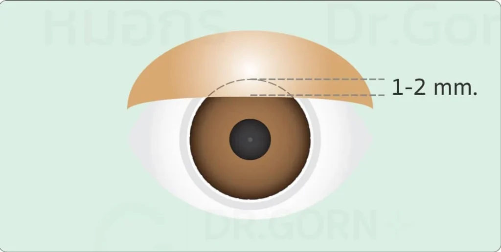
Mild Ptosis
The upper eyelid sits slightly lower than expected and covers more of the upper cornea.
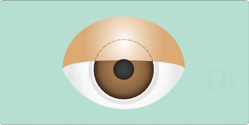
Moderate Ptosis
The upper eyelid descends further and may partially cover the pupil.
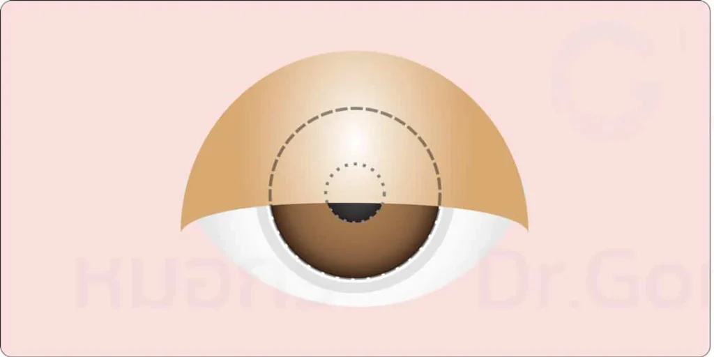
Severe Ptosis
The upper eyelid descends substantially and may cover a large portion of the pupil, interfering with vision.
The effects on daily activities and quality of life depend on the severity and underlying cause of the condition.
What Signs May Suggest Ptosis?
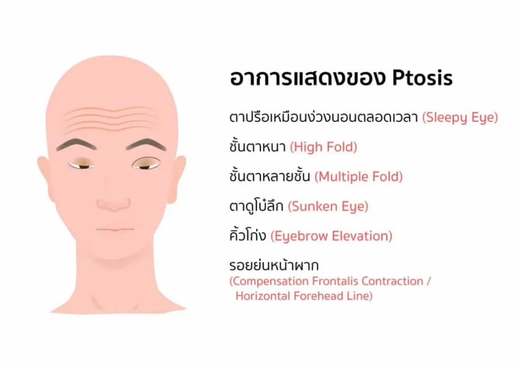
The eyes may appear partly closed, sleepy, or persistently tired. If only one eye is affected, the eyelid creases may look uneven. In some people, the drooping becomes more noticeable as the day progresses. Other possible features include a hollow upper eyelid area, a deep superior sulcus, or multiple eyelid folds.
More pronounced ptosis can reduce the upper visual field and make activities such as climbing stairs more difficult. Some people use eyelid tape or other products to help hold the eyelids open.
To compensate for drooping eyelids, the frontalis muscle may repeatedly contract to raise the eyebrows and eyelids. This can create a highly arched brow appearance and more prominent horizontal forehead lines.
Over time, these lines may remain visible even when the face is relaxed. Prolonged forehead muscle tension may also contribute to headaches. People who already experience migraines may notice an association between muscle tension and their symptoms.
Pseudoptosis: Conditions That Resemble True Ptosis
Pseudoptosis describes an appearance of eyelid drooping that is not caused by a primary problem with the upper eyelid lifting mechanism. Conditions that may produce this appearance include:
- Excess or loose eyelid skin, known as dermatochalasis, which is common with ageing.
- Downward misalignment of the eye, known as hypotropia.
- An unusually small eyeball relative to the eye socket, known as microphthalmia, or absence of the eyeball, known as anophthalmia.
- Eyelid conditions such as inflammation, tumours, cancer, excess fat, or enlargement of the orbicularis oculi muscle.
- Uneven eyelid changes associated with thyroid eye disease, such as retraction of the opposite upper eyelid.
- Protrusion of the opposite eye, which can make the other eye appear smaller by comparison.
If you are unsure whether you have true ptosis or pseudoptosis, consult a specialist before deciding to undergo double eyelid surgery.
What Causes True Ptosis?
1. Congenital Ptosis
Congenital ptosis is present from birth. It commonly involves incomplete development or abnormal structure of the levator muscle. Fibrotic tissue and abnormalities affecting other parts of the eyelid lifting structures may also reduce the ability to lift the upper eyelid.
Bringing childhood photographs to the consultation can help the doctor determine whether the condition was present early in life and assist with choosing an appropriate surgical approach.
2. Acquired Ptosis
Acquired ptosis develops after birth. Some forms progress gradually. Possible causes include:
- Age related changes: The levator aponeurosis may stretch, weaken, or become detached over time.
- Repeated stretching of the eyelid: Long term contact lens use or certain eye procedures involving prolonged eyelid manipulation may contribute to changes in the levator aponeurosis.
- Myasthenia gravis: This autoimmune disease affects communication between nerves and muscles. It may involve only the eye muscles or also affect other muscles, including those used for swallowing and breathing. Eyelid drooping and double vision may become more noticeable with sustained activity. Suspected cases require assessment by an ophthalmologist or neurologist because eye symptoms can be an early sign of a wider condition.
- Injury or previous surgery: Trauma to the levator muscle, levator aponeurosis, or both may cause ptosis. Previous double eyelid surgery may also contribute if these structures are injured. Correction requires careful assessment and surgical planning.
- Effects of botulinum toxin injections: Injections around the forehead, frown lines, or crow’s feet may cause eyelid drooping or make preexisting drooping more noticeable. Management may include waiting for the effects to wear off or using prescription eye drops in selected cases. The degree of improvement varies, and medication should be prescribed by a doctor.
What Happens If Someone With Ptosis Undergoes Conventional Double Eyelid Surgery?
If underlying ptosis is not recognised and addressed, conventional double eyelid surgery may leave the drooping unchanged or, in some cases, make it worse. Injury to the eyelid lifting structures or mechanical restrictions can affect their function.
Factors such as excessive bleeding, unusually high crease fixation, or excessive removal of the fat in front of the levator aponeurosis may complicate the outcome. Subsequent revision surgery can then become more difficult.
What Should You Do If You Suspect Ptosis?
- Consult a doctor to identify the cause. Ptosis is a sign that can arise from several different conditions, so treatment should address the underlying problem.
- Some causes require medical treatment before surgery is considered. Others may be suitable for surgery, while some require further investigation of other body systems.
- If the drooping is recent, including within the past six months, or you are unsure whether it has been present since birth, seek a specialist assessment before planning cosmetic eyelid surgery.
How Is Ptosis Caused by the Eyelid Lifting Structures Corrected?
When the problem involves the eyelid lifting muscle or its supporting attachments, ptosis correction surgery may be considered. The doctor selects an external or internal approach according to the examination findings. The expected result and the possibility of further correction depend on several factors:
- The type of ptosis: Congenital and acquired ptosis involve different tissue characteristics. Congenital cases with poor muscle function may be more complex to treat.
- The affected structure: The problem may involve the levator muscle, Müller’s muscle, the levator aponeurosis, or several components.
- Scarring from previous surgery: Extensive fibrosis can make revision surgery more difficult and limit the predictability of the result.
- Previous removal of orbital or preaponeurotic fat: Substantial fat removal may complicate revision and make tissue separation and management of recurrent scarring more challenging.
- The patient’s age.
- The timing of revision: For ptosis following surgery, the original article discusses either early correction within seven days or delayed correction after three to six months or longer. The appropriate timing must be decided individually by the treating specialist.