
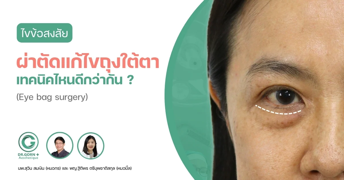
There is no single surgical technique that is best for every patient undergoing under-eye bag surgery (Lower Blepharoplasty). The most appropriate approach depends on the underlying problem, its severity, the supporting structure of the lower orbital rim and maxillary bone, the patient's age, and associated concerns such as lower eyelid laxity or deep under-eye hollows (Infraorbital Hollowness). To explain how surgeons may select a suitable technique, patients can be divided into three main groups.
Group 1: Prominent Under-Eye Bags Present Since Childhood or Adolescence
The first group includes patients who have had noticeable under-eye fat bags since childhood or adolescence, sometimes described as orbital fat hypertrophy or true eye bags. Many patients in this category are younger than 35 and do not have the following three associated conditions:
- No prominent natural dolly eyes (Pretarsal Orbicularis Oculi Hypertrophy). Patients who already have a pronounced pretarsal muscle fullness may notice that their natural dolly-eye appearance becomes more prominent after the underlying fat bags are reduced.
- No significant excess or sagging lower eyelid skin (Dermatochalasis).
- No significant under-eye hollowing (Infraorbital Hollowness).


A surgical technique that may be suitable for Group 1 is partial removal of the protruding orbital fat to reduce the appearance of under-eye bags and create a smoother lower eyelid contour. The surgeon may use a transconjunctival approach (Transconjunctival Orbital Fat Removal), in which the incision is made inside the lower eyelid. This technique avoids a visible external incision beneath the eyelashes. In appropriately selected patients, it may also reduce certain risks associated with external skin incisions. However, postoperative swelling and bruising can still occur, and recovery varies between individuals.
Steps Involved in Transconjunctival Under-Eye Bag Surgery
- The surgeon assesses the patient's lower eyelid anatomy and plans which orbital fat compartments require reduction and how much fat should be removed to achieve a smooth, balanced under-eye contour.
- The procedure is described in the clinic's original article as taking approximately 1–2 hours, often under local anesthesia. The duration depends on whether additional procedures are performed, such as autologous fat grafting beneath the eyes or fat transfer to the cheekbones and nasolabial folds.
- When sutures are required inside the eyelid, the surgeon may use absorbable sutures that do not usually need removal.
- According to the clinic's described follow-up schedule, postoperative examinations are arranged around days 7 and 30 to assess healing and recovery.
- Temporary swelling may occur after surgery and generally decreases during the first 1–2 weeks. The final contour may become clearer over approximately 1–3 months, although individual recovery times vary.


Group 2: Under-Eye Bags Caused by Orbital Septum Weakening, Muscle Laxity, and Aging
The second group includes patients whose under-eye bags develop or become more pronounced as the orbital septum and supporting eyelid tissues weaken with age. Changes in eyelid muscle support and connective tissue can allow orbital fat to protrude forward, making the under-eye bags more visible. Common characteristics include:
- Protruding orbital fat caused by weakening of the orbital septum and surrounding support structures. In more advanced cases, the fullness may extend toward the upper cheek, a condition associated with orbital fat herniation.
- Associated under-eye hollowness (Infraorbital Hollowness), which may result from naturally recessed lower orbital anatomy or age-related changes in the maxillary bone and surrounding soft tissues. The original article also mentions stress, smoking, and previous surgery as possible contributing factors, although these factors do not independently establish a diagnosis of bone retrusion.
- Prominent natural dolly-eye fullness resulting from the pretarsal portion of the orbicularis oculi muscle.
- Patients in this category are often older than approximately 35 years, although anatomy and tissue condition are more important than age alone.


A surgical technique that may be suitable for Group 2 is orbital fat transposition or redistribution. Instead of simply removing the protruding fat, the surgeon repositions selected orbital fat compartments to fill hollow areas beneath the eyes. This may be performed through a transconjunctival approach when significant skin removal is unnecessary. Alternatively, an external incision approximately 2 mm below the lower eyelid margin may be considered when excess skin and wrinkles also require treatment, or when additional midface lifting or correction of adjacent facial folds is planned.
Steps Involved in Under-Eye Fat Repositioning Surgery
- The surgeon examines and marks the lower eyelid and tear trough region to identify which fat compartments should be partially reduced and which should be repositioned. Important nerves, blood vessels, and other anatomical structures are also considered during surgical planning. The selected orbital fat may then be moved toward the recessed lower orbital rim or marked tear trough area and secured in the appropriate position.
- The original article describes an estimated surgical duration of 2–3 hours, potentially under local anesthesia. Actual time and anesthesia requirements depend on whether an internal or external incision is used and whether additional procedures are performed, such as:
- + Removal of excess lower eyelid skin.
- + Midface lifting or correction of sagging tissues and nasolabial folds.
- + Correction of lower eyelid ectropion or retraction resulting from previous surgery, or additional support for significantly lax lower eyelids.
- + Autologous fat grafting beneath the eyes, along the cheekbones, or around the nasolabial folds.
- The clinic's original postoperative schedule includes follow-up examinations on approximately days 5, 7, 14, and 30 to evaluate healing and the position of the treated tissues.
- When external fixation sutures are used to secure repositioned orbital fat, the surgeon may arrange their removal around postoperative day 5 or 7, depending on the technique and healing progress.
- The clinic advises avoiding contact lenses and strenuous exercise for approximately one month after surgery. Patients should follow their surgeon's individualized recovery instructions before resuming these activities.


Group 3: Patients with Both Prominent Under-Eye Bags and Deep Tear Trough Hollows
The third group includes patients who have characteristics of both Group 1 and Group 2. These patients commonly present with the following concerns:
- Excessively prominent or enlarged orbital fat bags beneath the eyes.
- Associated deep under-eye hollows or tear trough depressions. In these cases, the surgeon may consider combining partial orbital fat removal with orbital fat repositioning or redistribution during the same procedure to reduce excess fullness while preserving or restoring volume where needed.

As explained above, the most appropriate technique for under-eye bag surgery depends on the patient's individual anatomy and concerns. Anyone considering lower blepharoplasty should consult a qualified surgeon experienced in eyelid surgery for a thorough assessment. The surgeon can then recommend an approach that balances the intended cosmetic improvement with eyelid support, healing considerations, and potential complications. No surgical technique can guarantee a complication-free outcome.